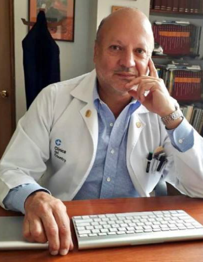
Ophthalmology studies the different diseases that can affect the eyes, dedicating itself to preventing, diagnosing, and treating diseases including surgery when treating different conditions.

Certified ophthalmology surgeon with a subspecialty in ocular oncology, orbit, cornea, ocular surface, and refractive surgery. More than 25 years of experience in which I have developed a prestigious full-time service in clinical-surgical practice.
I am the director and associate professor of ophthalmology in the Orbit and Oncology Unit of the National Institute of Ophthalmology. I am also the founder and director of the Oculoplastic, Orbit, and Oncology Scholarship Program at the Military Central Hospital affiliated with the Armed Forces of Nueva Granada University and Calle Centro Oftalmológico in Bogotá, Colombia.
I have been the Postgraduate Medical Director at the San Rafael University Hospital and Clinic for 7 years and I am a specialist in medical education, epidemiology, and critical analysis of scientific literature from the Universidad de Los Andes and medical ethics from the Universidad El Bosque.
I did my residency at the Hospital y Clínica de la Universidad de San Rafael affiliated with the Nueva Granada Military University in Bogotá, Colombia and in ultrasound and ophthalmic photography at the Bascon Palmer Eye Institute in Miami to later do a residency in Ocular and Orbital Oncology at Wills Eye Hospital in Thomas Jefferson University of Philadelphia, United States.
I am an official reviewer for the journals of Ophthalmology, Eye, BMJ, AJO, Orbit (in the field of Oncology and Orbit), international consultant on Orbit tumors for the PAAO (Pan American Association of Ophthalmology), and editorial editor of Franja Ocular for the past six years.
For the past two years I have been the Founder and Director of the Latin American Program of the Ocular-Orbit Oncology Fellowship sponsored by the Eye Cancer Foundation of New York and a member of the Foundation's scientific advisory committee. I have given many international conferences in Latin America, North America and Europe and I have received prestigious awards including JCI (Award for 10 Young Creative Entrepreneurs 1996), Ocular Tumos Board (AAO) 2001, 100 Best Doctors in Latin America (For 10 years).
The areas of interest and research of my clinic include Ocular and Orbital Oncology, Immunotherapy, Orbital Trauma, Multidisciplinary Orbital Reconstruction Surgery, Refractive Surgery, Aesthetic and Non-Aesthetic use of Botulinum Toxin (Botox) and Cataract Surgery.
I inherited my passion for medicine, ophthalmology, and surgery from my father, who was also a professor of ophthalmology with whom I had the privilege of working for several years.
Languages: Spanish, English, Portuguese and Italian.
I do this procedure using premium intraocular lenses of the latest technology, monofocal, trifocal, etc.
The cataract operation consists of the extraction of the part of the lens that is opacified to restore vision to the eye. In general, there is a tendency to replace it with an artificial lens that is placed in the same place as the original crystalline lens (intraocular lens) or restoring the vision that had been lost because of cataracts. Just like when a glass of glasses is damaged, we change it for a new one, when a cataract occurs, we must change the lens or “glass” that we have inside.
This is why the treatment of a cataract is exclusively surgical. To do this, we extract the opaque lens, either in its entirety or by opening its capsule and extracting the inner part or nucleus, the latter being the usual form of intervention. To compensate for the loss of the eye’s natural lens, a synthetic material lens is inserted, known as an intraocular lens, thus replacing the crystalline lens, which allows for better visual recovery. For the extraction of the nucleus of the crystalline lens we have two options, either it can be extracted in its entirety, which requires making a large incision to facilitate its exit, or it can be removed directly inside the eye using ultrasound or laser.
In the first case, the technique is called extracapsular lens extraction, and its use is currently infrequent. When we directly remove the cataract inside the eye, the technique is called phacoemulsification, this being the standard form in cataract surgery since it requires a minimal incision and allows faster recovery.
The LASIK technique (acronym in English for “Laser in situ Keratomileusis”) is currently the most widespread due to its safety and efficacy. It consists of modifying the shape of the cornea (keratomileúsis: from the Greek, kerato: cornea, and mileúsis: to sculpt) by applying the excimer laser inside it.
Previously, a thin layer of corneal tissue has been raised, which is later repositioned and adheres without the need for stitches. The correction of the refractive error is carried out in this way, with minimal discomfort for the patient and a very fast recovery.
Anesthesia for this surgical technique is topical (with eye drops) and postoperative eye bandage is not necessary. The precision and safety of the LASIK technique make it the surgical procedure of choice for most refractive errors.
Lasik:
The LASIK technique (acronym in English for “Laser in situ Keratomileusis”) is currently the most widespread due to its safety and efficacy. It consists of modifying the shape of the cornea (keratomileúsis: from the Greek, kerato: cornea, and mileúsis: to sculpt) by applying the excimer laser inside it.
Previously, a thin layer of corneal tissue has been raised, which is later repositioned and adheres without the need for stitches. The correction of the refractive error is carried out in this way, with minimal discomfort for the patient and a very fast recovery.
Anesthesia for this surgical technique is topical (with eye drops) and postoperative eye bandage is not necessary. The precision and safety of the LASIK technique make it the surgical procedure of choice for most refractive errors.
Lasek:
The term LASEK (Laser-Assisted Subepithelial Keratectomy) means Laser-Assisted Subepithelial Keratectomy and is a technique that could be considered a combination between PRK and LASIK.
What you can expect during the procedure:
• Before treatment, you may be given medicine to help you relax.
• You will be given eye drops to numb your eyes.
• A device is used to keep your eyes open.
• An alcohol-soaked instrument will be briefly placed on your cornea. The surgeon will then push the softened epithelium back to expose the inner tissue of the cornea.
• The surgeon will use a computer-guided excimer laser to reshape the cornea. The laser treatment lasts 10–90 seconds.
• The epithelium redraws over the cornea.
• For a few days you will have to wear a contact lens as a bandage to protect your cornea while it heals.
Upper and lower eyelid reconstruction:
Eyelid reconstruction is a surgical procedure used to correct eyelid defects that result from the surgical resection of tumors, trauma, or congenital anomalies such as a coloboma. Reconstruction of the eyelids due to surgical resection of malignancies, such as skin cancers removed by Mohs micrographic surgery, requires additional consideration.
The restoration of the upper eyelid is much more complicated than that of the lower eyelid. Careful deliberation is necessary for the approach to reconstruction since the repair is highly dependent on the location and extent of the defect.
The eyelids fulfill essential functions for the face. In addition to providing a cosmetic appearance, the eyelid mechanically protects the cornea and the eyeball. In addition, meibomian glands in the tarsus produce lipids that, by contracting the orbicularis oculi tarsi, stabilize the tear film to prevent dry eyes.
Eyelid reconstruction is necessary whenever there is an eyelid defect due to a combination of possible cosmetic and functional deficits, as mentioned above. Depending on the results of the surgery, revision surgery may be necessary
Upper blepharoplasty:
The eyelids can lose their natural position for different reasons, ranging from changes at birth to their senile evolution. In these conditions, the eyelids can droop, which is known as palpebral ptosis, they can remain open, which is an ectropion, or they can remain inward, a condition known as entropion. In these situations, various discomforts are produced, either by tearing in ectropion, by rubbing against the eyelashes in entropion or loss of field of vision in palpebral ptosis.
When the eyelids lose their stability and function, the oculoplastic surgeon must perform the appropriate anatomical correction to re-establish the original conditions of the eyelid by reinserting the palpebral tendons or advancing the muscles that have lost their strength.
These surgeries have, in addition to a functional and restorative purpose, a cosmetic approach as they are made through invisible or hidden incisions.
Lower blepharoplasty:
Cosmetic eyelid surgery is scientifically called blepharoplasty. This cosmetic surgery allows you to correct the fatigued or aged appearance in the look due to changes around the eyes.
Cosmetic eyelid surgery is performed directly on the eyelids without touching the eyeball. With advancing age, the skin of the eyelid stretches, the muscles weaken, and fat accumulates around the eyes, causing “bags” to form at the top and bottom.
This cosmetic eyelid surgery “blepharoplasty” removes excess skin, fatty bags, if any, and part of the orbicularis muscle, if necessary, improving the appearance of tired eyes, refreshing them, and rejuvenating them.
Uveal melanoma: An intraocular melanoma is one of the most common forms of eye cancer. It can develop in the iris, the ciliary body (region of the eye behind the iris that produces aqueous humor), or in the choroid vascular at the back of the eye. This form of eye cancer occurs most often in adults 60 years of age and older.
Intraocular lymphoma: This form of cancer is a rare form of lymphoma that begins in the eyeball.
Eyelid tumors: The most common form of eyelid cancer is a skin cancer called basal cell carcinoma. There are also other types of eyelid tumors, such as malignant melanoma, sebaceous cell carcinoma, and squamous cell carcinoma. Most of these tumors can be removed with surgery.
Conjunctival tumors: Lymphomas, melanomas, and squamous cell carcinomas are tumors that grow on the surface of the eye.
Lacrimal gland tumors: This type of tumor grows in the lacrimal glands.
Retinoblastoma: This form of cancer is a cancer of the retina, the light-sensitive tissue of the eye. Retinoblastoma, the most common childhood eye cancer, usually develops in children younger than 5 years. In almost a third of cases, retinoblastoma occurs in both eyes due to a mutation in the RB1 gene. Parents often notice leukocoria, or white color in the pupil, as the first sign, which results from retinoblastoma tumors
What are they:
Orbital tumors are abnormal growths of tissue in the structures surrounding the eye. These lesions may be benign or malignant and may arise primarily from the orbit or may spread (metastasize) from elsewhere in the body.
The most common types of orbital tumors vary considerably by age, but include cysts, vascular lesions arising from blood vessels, lymphomas, neurogenic tumors arising from nerves, and secondary tumors either metastatic or spread directly from the surrounding sinuses or the cranium.
Treatment:
There are a variety of treatment options for these tumors, and the modality used depends on the type of tumor. Whenever possible, these lesions are removed by careful surgical techniques.
However, not all tumors require surgical removal, and for some, radiation, chemotherapy, or immunotherapy may be the indicated form of treatment.
Orbital fractures are breaks in any of the bones that surround the eye area (also known as the orbit or eye socket). These fractures are almost always the result of blunt force trauma, either from an accident or from sports. Orbital fractures can cause a multitude of problems depending on where they are located and what other associated injuries may be present. Because of this, detailed ophthalmologic evaluation is paramount to see which fractures may require correction to restore normal visual function.
Types of orbital fractures:
Orbital fractures can be classified into three different types:
• Blowout Fracture: Most common, a blowout fracture occurs in the eye socket along the floor or inner wall near the nose and is often caused by something hitting the eye with force, such as a tennis ball or racquetball. These fractures may be asymptomatic and may be seen or cause double vision problems, or a change in the position of the eyeball, and require surgical repair.
• Orbital Rim Fracture: These fractures are found at the outer edges of the eye socket and require a great deal of force to inflict. Orbital rim fractures are often the result of motor vehicle accidents, accompany other head and facial injuries, and may present as an irregular contour along the rim of the eye socket.
• Compound Fractures: Trauma to the midface can result in a combination of fractures including the orbital rim, floor, and cheek, and may be referred to as a tripod fracture or ZMC (zygomaticomaxillary complex). This can affect both the eye socket and the maxilla or upper jaw, resulting in abnormalities when biting or chewing. Other associations of compound fractures include the bones of the nose, the cranial vault (chamber that contains the brain), and the base of the skull.
If the fracture has affected the movement, function, or location of the eye, reconstructive surgery may be needed.
Dejanos tu correo y recibe actualización de todos nuestros servicios y noticias.